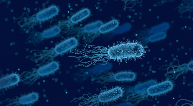
A desktop diagnosis tool that detects the presence of harmful bacteria in a blood sample in a matter of hours instead of days has been developed by engineers at the University of California San Diego. This was made possible by a combination of proprietary chemistry, innovative electrical engineering and high-end imaging and analysis techniques powered by machine learning. The details of the team’s work are published in Scientific Reports.
Researchers melted bacterial DNA in 20,000 extremely small simultaneous reactions to identify low levels of harmful bacteria among a large number of human blood cells. One drop of rain contains hundreds of thousands of picoliter whereas each of these reactions contained only 20 picoliters. As DNA comes apart during melting, each one has a specific signature. Researchers can use machine learning to determine which types of DNA appear in blood samples as the melting process is imaged and analyzed. The system accurately identified DNA sequences from bacteria causing food-borne illnesses and pneumonia in less than 4 hours in 99% of the time.
Stephanie Fraley, a professor of bioengineering at the Jacobs School of Engineering at UC San Diego and the paper’s lead author said that analyzing these many reactions at the same time at this small a scale has never before been attempted. This is a much more holistic approach. Currently, the methods being used to detect and identify bacteria relies on cultures that can take days and is too long to provide physicians with an effective and timely diagnosis tool before prescribing antibiotics.
One milliliter of blood is inoculated with Listeria monocytogenes, a foodborne bacterium that causes 260 deaths a year in the U.S., and Streptococcus pneumonia, which causes everything from sinus infections to pneumonia and meningitis.
All DNA was isolated from the blood sample and then placed on a digital chip that allowed each piece to individually multiply in its own small reaction. Each well containing DNA in the chip was only 20 picoliters in volume and researchers used a proprietary mix of chemicals subject to a provisional patent. The chip with the amplified DNA was placed in an innovative high-throughput microscope that the team had designed and then heated in increments of 0.2 degrees Celsius, causing it to melt at temperatures between 50 and 90 degrees, or about 120 to 190 degrees Fahrenheit.
The bonds holding the DNA strands together break as the DNA double-helix melts. The bonds have different strengths and that changes the way the strands unwind from each other creating a unique sequence-dependent fingerprint that can be detected using a special dye. The dye used causes the unwinding process to give off fluorescent light that creates what the researchers call a melting curve or unique signature for each type of bacteria. They were then able to capture the bacteria’s melting curves when imaging the melting process with the high-throughput microscope. The curves were then analyzed with a machine learning algorithm they developed.
After this process shrinking the size of the system was done so that it could be more easily deployed in clinics and physicians’ offices. They want to add to the system the capability to date fungal and viral pathogens, as well as genes for antibiotic resistance. They hope to further validate the results on patient samples and make this available to physicians in the next 5 years.
Many treatments fall under the umbrella of Complementary and Alternative Medicine or CAM. Some of the most commonly used CAM therapies include: Acupuncture Chiropractic Food counseling Herbalism Massa...
For many centuries, Thanksgiving has been considered a national holiday in the U.S. and Canada to celebrate the harvest and other blessings of the preceding year. The traditions behind it have evolved...
Polls in the United States and Europe revealed that half the population are more afraid of cancer than any other disease. Cancer is, after all, a life-altering event that can trigger a rollercoaster o...